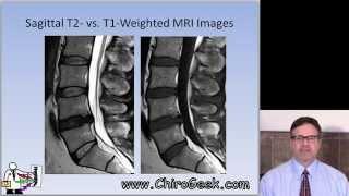
Dr. Gillard lectures on How to Read Your Lumbar MRI
Комментарии:

Not a student, just a patient trying to understand my mri.
This was 9 years ago. Do you have a link to any updated versions
Thanks Dr.
Will subscribe for sure

Excellent informative lecture thank you very much.
Ответить
In the parasagital view the view is t1 but it written t2 by mistake I think.
Ответить
An outstanding explanation, Thank you so much, professor🤍
Ответить
Wonderful Overview.. Thank you so much!!
Ответить
why do mri scan not scan the bottom nerves s2 to s5 ?!
Ответить
Dear Dr gillard, lovely explanation .of MRI of lumbar spine.
Ответить
Thank you kindly! A wealth of information indeed.
Ответить
much appreciated, usefull information, thanks doc
Ответить
On my mri the spine doctor said one of my disc's is all black in the image meaning it's very bad because they are supposed to look white like marshmallows
Ответить
I highly recommend you second guess your radiologist 😂, as a matter of fact I also recommend doing the exact opposite of what the prescription bottle says to do, take 1 every 4 hours…NOPE TAKE 2 every hour for 4 hours 😂
Ответить
Well explained Doctor
Ответить
Thank you 🇺🇸
Ответить
Please doctor I need to speak with u
I want to show you my MRI 7 years I suffer I didit found solution my age now 25 years old

Thanks for the amazing, and heretofore totally mysterious, explanation of lumbar MRIs. I have a long way to go to get close to full understanding, but at least it's starting to make sense as I view my own MRI. Cheers.
Ответить
You have made MRI reading as simple as if an X-ray reading. Great
Ответить
Really appreciate you taking the time to share your knowledge.
Ответить
نريد ترجمه باللغه العربيه
Ответить
Great talk ....Thank you
Ответить
thanks for your videos douglas, very informative. heres to hoping myself and many others get pain relief one day, small bulge club. 👎
Ответить
Thanks. Amazing at how many shade tree radiologists we have out there!
Ответить
Thanks it really helped me understand viewing MRI. 👍😊
Ответить
Fig 7 picture, it that a wrong label of Left SAP L4 vs IAP L5? tks
Ответить
This is great for me.. I passed 2 boards in radiology but before MRI. After 40 years and 1000s of patients. It's time to get this down.. Thank you very much Bless you Doctor🙏
Ответить
After countless googles on how to read an MRI of the lumbar, your page came up on my homepage randomly. This is by far the simplest and most informative description ( to a layman ) of how to read an MRI. Great video!
Ответить
Thank you for the lesson Prof. Dr. Douglas Gillard🙏🙏🙏. This gives me broader insight of MRI as I am not a radiologist.
Ответить
So great
Ответить
Enjoyed, I have a fusion L5 S1, due to the fusion S1, S2 have been damaged by scar tissue, making for pain.
Ответить
I am 25 years old, I worked a few months, I was sitting all day in uncomfortable positions without recharging for hours and from then on I have very annoying muscle pains that I feel more at night, I feel them on the left and right side of my entire back I am I'm sure the pain is in the muscles as if they were tense a lot, I already went to the chiropractor and he didn't help me at all, he just broke the bones of my spine, he didn't do anything similar to what this man does, I'm tired already having these pains , of not sleeping well, doctor if you see this comment please tell me what I can do😓
Ответить
Excellent video. After staring at the spine for so long and imagining how everything is, I believe you need a skeleton to really get it down. Takes a while the other way. You need to have a 3 d mind.
Ответить
Dr you talk so clear and perfect that it sound like your eating the most expensive meal and you are enjoying every single bite.
thank you so much I am not a doctor but I love and enjoy learning new things.

CHIROPRACTOR MESSED ME UP BAD BAD BAD
Ответить
Awesome lecture Sir..
Ответить
please we want your clear conception lectures on thoracic and especially cervical spine which is very difficult to interpreto
Ответить
Hola, buenas tardes. Tienen el video en español por favor. Gracias!!
Ответить
Thanks a lot.. Doctor.. Can I send you a ct.. I need ypur opinion.. Please.
Ответить
I m grateful for you dear, Alot
Ответить
please reply on your email i have sent one
Ответить
Thank you. Easy to understand when You speak it.
Ответить
Awesome video! Tyvm
Ответить
Would appreciate some help regarding C4-C5 cord atrophy with compressive myelomalacia and what this means exactly?
Appreciate any advice on suggestions💯
Coronal localizer images of series 1 provides partial visualization of the upper
portion of a thoracic levoscoliosis.
There is straightening of normal cervical lordosis. No spondylolisthesis is
visualized.
There is no evidence of fracture, osteomyelitis or osseous neoplasm.
Postoperative changes are again noted is provided below.
1. Patient has undergone anterior cervical discectomy, instrumentation and
fusion from C3 to C6.
2. There has been partial laminectomy at C3-C4 and C4-C5.
3. Posterior instrumentation artifact is visualized from C3 to C6.
No postoperative complication is visualized.
The atlanto-occipital and atlanto-axial articulations are unremarkable in MR
appearance.
The visualized intracranial structures are unremarkable in non-contrast MR
appearance.
The spinal cord is visualized to the T5 level. Again noted is myelomalacia and
cord atrophy at the C4-C5 level..
C2-C3: Minimal spondylosis. Minimal right facet arthrosis. Minimal disc bulge.
C3-C4: Anterior fusion. Partial laminectomy and posterior instrumentation.
Minimal left uncovertebral joint spurring.
C4-C5: Anterior fusion. Partial laminectomy and posterior instrumentation.
C5-C6: Anterior fusion. Posterior instrumentation.
C6-C7: Moderate-to-severe spondylosis. Severe left facet arthrosis. Small
left-sided disc-ridge complex, without compression of the left C7 nerve root.
See axial series 6, image 17.
C7-T1: Moderate spondylosis. Facet arthrosis, moderate on the right mild on the
left. Small right-sided disc protrusion without surrounding mass effect,
sagittal series 2, image 9.
The upper thoracic spine is visualized to the T5 level on sagittal sequences.
There is minimal spondylosis from T3 to T5. There are small central and
right-sided disc protrusions at T3-T4 and T4-T5, mildly effacing the ventral
thecal sac, sagittal series 2, image 7-10, unchanged compared to sagittal images
11 of series 2 from the June 2020 MR study.
There is mild postinflammatory mucoperiosteal thickening in both maxillary
antra.
Impression:
Straightening of normal cervical lordosis.
Postoperative changes as detailed above, without postoperative complication.
Chronic compressive myelomalacia and cord atrophy at the C4-C5 level, unchanged
compared to preoperative studies.
Spondylosis and facet arthrosis as noted.
Small left-sided disc-ridge complex at C6-C7, without compression of the left C7
nerve root.
Small right-sided disc protrusion at C7-T1, without significant mass effect.
Small central and right-sided disc protrusions at T3-T4 and T4-T5, unchanged
compared to MR images from June 2020.

I have spinal stenosis and find your information very valuable. Trying to decide between a versiflex or PRP. Thanks.
Ответить
Hello.
Just a quick question.
If the radiologist does not put in the correct weight and height. Will that influence with the readable of the MRI?
He programmed a hight of 170 cm and a weight of 70kg. And I am 180cm and 105kg.
I also asked if the cut wil be 3mm cut. And he used 4mm cut.
I followed your video about your preferred MRI reader.
Al images of the lumbar are a kind of blurry compared with an other older series of my thorax and cervical spine.
Your suggestions are appreciated.
Best regards Arny Koch retired and 63

![How to Use Projects Bones in FL Studio 20 [Copy and Paste Between Projects] How to Use Projects Bones in FL Studio 20 [Copy and Paste Between Projects]](https://invideo.cc/img/upload/WWZSQUJsUk11b1E.jpg)


