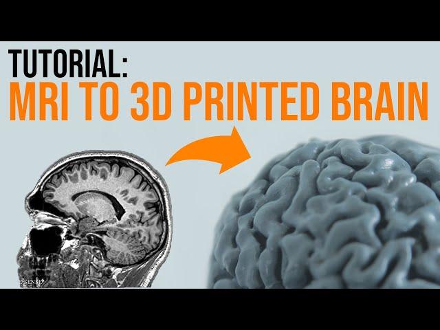
How To 3D Print Your Brain In A Few Simple Steps - TUTORIAL 2022
Комментарии:

What if we wanted to print also the inside structures in order to create an assemblable model?
Ответить
Great video! Thank you👌
Ответить
So I followed the steps but was a little concerned when I saw my brain in 3d editor looking like a literal sponge, no folds in the brain just a sponge, do you have any fixes for that or is it the MRI issue?
Ответить
that's awesome! great job
Ответить
DUDE! You must 3D print a Jello brain mold! Imagine at your next party, serving your brain laced with fruits, sweets, maybe nuts, and of course "happy" ingredients. Lmao! Also 3D print your skull, as a serving dish for the brain. Definitely.
Ответить
Thats a great video!
I just followed your instruction, but wen it comes to the "Make Solid" step in Meshmixer, than I just get a transparent brain and whenI accept it, its just gone. Do you have any idea what I did wrong?

Your tutorial was the best one, thank you!!!
Ответить
When I am looking at my mri images on the 3 different planes only 1 is high res and the other 2 are not. I can click on a different image and then a different image out of the 3 planes is high res. In the example you show all 3 planes are high res. Do all 3 need to be high res or is there a way for me to take the high res of each image and combine them so I have all 3 planes high res?
Ответить
Nice tutorial, but I don't like how you did 3D slicing part:
Making cylindrical voids adds more paths for printer, you should just change infill amount.
Splitting onto 3 parts is kinda pointless, just make it whole thing. Or splice it by brain halfs. Or make it smaller.

Great video, super helpful!
I'm also struggling with lack of resolution in the resulting model. I can't see any individual folds the way that you have in your resulting model. I've played with the thresholding a lot and it hasn't helped.
What size do you consider to be high res? The 'series' I am using is 512x512 size, with 176 as the 'count' column.

Great video, my MRI scan has many views, and I'm unsure which to pick to do this. The ones I have picked so far seem to be low resolution for the side and rear, high res on the top.
Ответить
Hi, How do I get in touch with you? My son has had a corpus callosotomy performed in 2011. It now appears on the MRI that 2 connections have remained resulting in sever epilepsy again. The neuro surgeon suggested a 3D printed version to inspect each part to ascertain if further surgery will be beneficial... I know - tough one...
Ответить
I am using data from a clinical study. Amazing detail and an amazing tutorial so far.
Ответить
This is the most comprehensive tutorial I have found online for this, thank you!!!
Quick question though, whenever I pull my files into Slicer, the back of my brain is always less detailed than the top front. Do you have any suggestions to fix this? All the tissue folds basically combine into one semi-flat semi-bumpy area. Scrubbing through the files in the DICOM viewer shows that the rear area of my scans looks identical to the rest. So I don't know why I'm having this issue. If I change the threshold it's either too flat on the back, or if I adjust the threshold so the back looks good, the front top becomes way too sharp and the folds aren't even close to one another and it looks like veins instead of tissue folds.
Is there any way to apply a gradient to the threshold? Any advice is appreciated!!!

Thank you for this, exactly what I was looking for! Unfortunately when I press apply no 3d model appears in the viewer.
Ответить
Thank you! I participate in the LE TBI study at UWMC in Seattle and I get MRI scans... so soon maybe i can print my brain
Ответить
I followed your tutorial and shared the resulting print on reddit, and now tons of people are asking how I did it so I’m just linking them to your video
Ответить
My MRI images are in JPG, I never downloaded a DICOM file and I doubt the records are easily accessible from being years ago, and can't be stuffed contacting them for it. I could spend time learning how to add that into 3D Slicer to be usable, or convert it to DICOM, but I just learnt that the set of images is missing the Coronal axis. Unless you tell me otherwise, this seems important for the process, and I have stopped.
Ответить
thankyou!
Ответить
hello is there a tool that merge 3 different series? because when i open one series axial is good but others bad and in the other coronal is good and in the other sagittal is good i want to merge good ones to use in segment editor to make 3 model
Ответить
They could do an AI tool to automate the conversion from MRI to 3d model. That would be awesome. Thanks for the video!
Ответить
How to check vein that is going above trigeminal nerve into 3d to see contact is there or not??
Ответить
Me ending up with an empty .STL file😢
Ответить
Is there a way to do this but for the bones instead????
Ответить
Bro, i don't mean to be a party pooper, but you printed your brain with -45 IQ points...you cut away a lot of gray matter hahahaha (that segmentation was too aggresive)
Ответить
Could you make your skull instead of your brain, using the same scans? That would be very cool.
Ответить
I have a different series for each angle (axial, sag, coronal) and combining them into one image in 3D slicer has been a nightmare
Ответить
This is a great tutorial. My 3d model in Slicer isn't as defined as yours. Is that because of the actual file sizes of the MRI or is it just settings in the software? I'm wondering if the images on my CD from the radiologist aren't hi-res enough.
Ответить
Great tutorial! I did something similar back in 2014, and it was a massive pain. This looks so much easier in comparison! Thanks for sharing!
Ответить
What about from CD? How do you get it on the computer to begin with?
Ответить
About how long did the design (pre-print) process take? Hard to tell since you had to edit and clean up for YT (not complaining, just trying to understand a reasonable expectation)
Ответить
Thanks for the tutorial! Do you know how i could isolate and print the Skull by itself?
Ответить
I have a problem with my source data. I have various sets, each of them for example the axial one is high quality only on the axial plane, and low quality on the other two planes. Same thing for the other two sets. But it seems like in the input volume I can only select one set at time. Is there any way to make it work for my case? :\
Ответить
Really nice and cool tutorial, Thank you!!
Ответить


